Neck Muscle Diagram ~ 1 The Anatomy And Physiology Of The Neck Ento Key. The muscles of the neck anatomical chart shows in beautiful detail the many anterior, posterior, inferior and lateral views of every muscle that makes up the matrix of support for our skull and brain. Head and neck muscle diagram. Thank you for your support. The next life study seated female figure, shows the upper part of the pectoralis major positioned flat against the rib cage, with. The anterior and middle scalenes originate from the transverse processes of certain cervical.
The muscles of the neck run from the base of the skull to the upper back and work together to bend the head and assist in breathing. Many in the neck help to stabilize or move the head. The hyoid bone and other such structural components require the cooperation of neck muscles as well. Posted on december 4, 2018december 3, 2018. The three scalene muscles are found forming the floor of the posterior triangle.
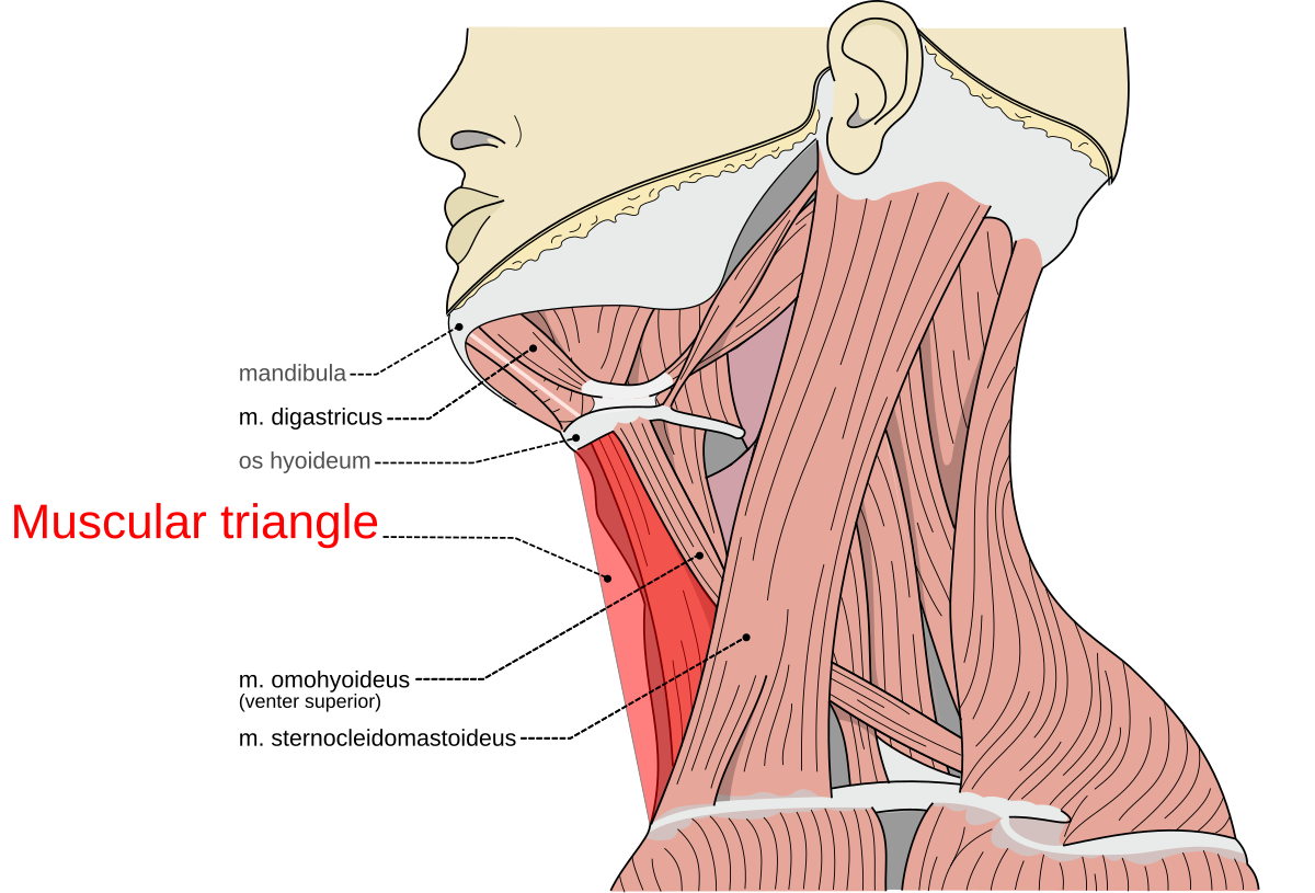
Muscular Triangle Wikipedia from upload.wikimedia.org Many in the neck help to stabilize or move the head. Neck neck muscle anatomy muscle diagram inspirational medical. The next life study seated female figure, shows the upper part of the pectoralis major positioned flat against the rib cage, with. Head and neck muscles diagram so many muscles that cause migraines arm neck shoulders and back templates. Each side of the neck contains two triangular sections created by the major deep muscles. 9 видео 363 062 просмотра обновлен 5 февр. Related posts of anatomy of neck muscles diagram. The muscles of the neck anatomical chart shows in beautiful detail the many anterior, posterior, inferior and lateral views of every muscle that makes up the matrix of support for our skull and brain.
Head and neck muscles diagram so many muscles that cause migraines arm neck shoulders and back templates.
Head and neck muscles diagram so many muscles that cause migraines arm neck shoulders and back templates. Neck anatomy pictures bones muscles nerves. 9 видео 363 062 просмотра обновлен 5 февр. There are three sections for you to practice. Automatic on off switch for water pump circuit diagram. Human muscle system, the muscles of the human body that work the skeletal system, that are under voluntary control, and that are concerned with movement, posture, and balance. Located in the front of the neck, the anterior triangle includes four smaller triangles. This diagram depicts head neck muscle diagram. Exercise 15 gross anatomy of the muscular system. The splenius capitis and the splenius cervicis are neck extensors. Human muscle system, the muscles of the human body that work the skeletal system, that are under voluntary control, and that are concerned with movement, posture, and balance. This is a table of skeletal muscles of the human anatomy. The muscles of the neck anatomical chart shows in beautiful detail the many anterior, posterior, inferior and lateral views of every muscle that makes up the matrix of support for our skull and brain.
Anatomy of neck muscles diagram. Er diagram to relational schema. The muscles of the neck anatomical chart shows in beautiful detail the many anterior, posterior, inferior and lateral views of every muscle that makes up the matrix of support for our skull and brain. Neck diagram of muscles, arteries, and skeleton. Human anatomy diagrams show internal organs, cells, systems, conditions, symptoms and sickness information and/or tips for healthy living.
Axial Muscles Of The Head Neck And Back Anatomy And Physiology from s3-us-west-2.amazonaws.com They move the head in every direction, pulling the skull and jaw towards the shoulders, spine, and scapula. There are three sections for you to practice. Posted on december 4, 2018december 3, 2018. Advertisements help pay for this website. Muscles diagram human head and neck muscles diagram 9 out of 10 based on 70 ratings. Er diagram to relational schema. The hyoid bone and other such structural components require the cooperation of neck muscles as well. 5 best images of printable college anatomy worksheets.
There are three sections for you to practice.
The muscles of the neck can be divided into groups according to their location. In anatomy, the temporal muscle, also known as the temporalis, is one of the muscles of mastication. The three scalene muscles are found forming the floor of the posterior triangle. Located in the front of the neck, the anterior triangle includes four smaller triangles. Posted on december 4, 2018december 3, 2018. Neck neck muscle anatomy muscle diagram inspirational medical. Neck anatomy pictures bones muscles nerves. This is a table of skeletal muscles of the human anatomy. The stylohyoid muscle is this muscle here, which connects from the styloid process of the skull to the lateral surface of the hyoid bone those are the four suprahyoid muscles in the anterior triangle of the neck. For more anatomy content please follow us and visit our website: Er diagram to relational schema. Head and neck muscles diagram anatomy note we are pleased to provide you with the picture named head and neck muscles diagram we hope this picture head and neck muscles diagram can help you and satisfies your requirements anatomynote found head and neck muscles diagram from plenty. Head and neck muscle diagram.
Distinguish the muscles that developed on the basis of the first (mandibular) and second (hyoid) visceral, gill arches, and muscles that developed from the ventral divisions of the myotomes. The stylohyoid muscle is this muscle here, which connects from the styloid process of the skull to the lateral surface of the hyoid bone those are the four suprahyoid muscles in the anterior triangle of the neck. The sternocleidomastoid muscle separates the sections, known as the anterior and posterior triangles. Er diagram to relational schema. The main functions of the neck muscles are to permit movements of the neck or head and to provide structural support of the head.
Splenius Capitis Muscle from www.getbodysmart.com The muscles of the neck anatomical chart shows in beautiful detail the many anterior, posterior, inferior and lateral views of every muscle that makes up the matrix of support for our skull and brain. The stylohyoid muscle is this muscle here, which connects from the styloid process of the skull to the lateral surface of the hyoid bone those are the four suprahyoid muscles in the anterior triangle of the neck. Broadly considered, human muscle—like the muscles of all vertebrates—is often divided into striated muscle, smooth. Related posts of anatomy of neck muscles diagram. There are three sections for you to practice. Muscle identification muscle actions and muscle origins and insertions. Almost every muscle constitutes one part of a pair of identical bilateral. Artists artwork backmuscles body comic comics dynamic illustration muscle reference studies stylized superhero sternocleidomastoid neckmuscles art anatomy drawing muscles neck trapezius.
The splenius capitis and the splenius cervicis are neck extensors.
The three scalene muscles are found forming the floor of the posterior triangle. The stylohyoid muscle is this muscle here, which connects from the styloid process of the skull to the lateral surface of the hyoid bone those are the four suprahyoid muscles in the anterior triangle of the neck. Exercise 15 gross anatomy of the muscular system. The quizzes below each include 15 multiple choice identification questions related to the muscles of the head and neck. Head and neck muscle diagram. The muscles of the neck are present in four main groups. Posted on december 4, 2018december 3, 2018. We hope this picture head and neck muscles diagram can help you study and research. Working in pairs on the left and. Each side of the neck contains two triangular sections created by the major deep muscles. In anatomy, the temporal muscle, also known as the temporalis, is one of the muscles of mastication. It runs superolaterally from the sternum and clavicle to the lateral surface of the mastoid process. Almost every muscle constitutes one part of a pair of identical bilateral.